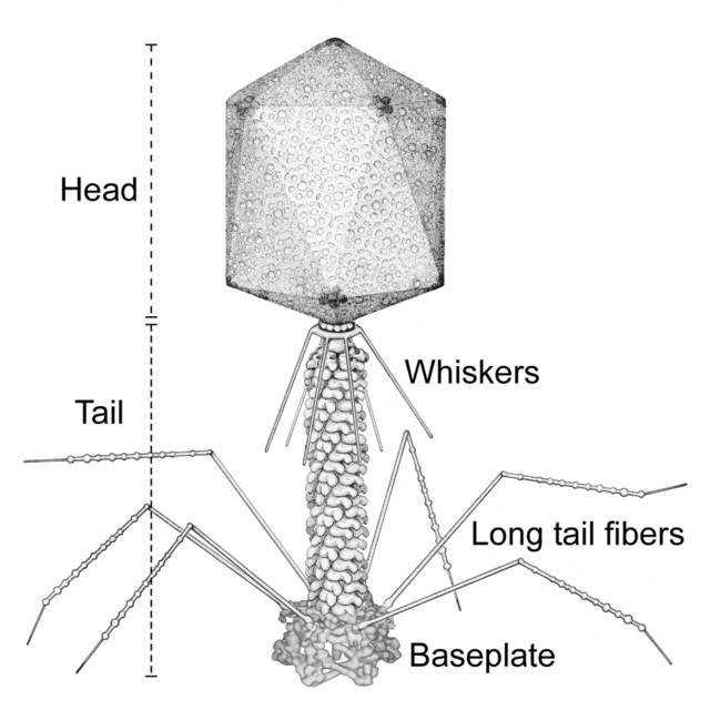
Scematic of a T4 bacteriophage (Purdue)

I knew that in the end, the proteins that make up the organelles, walls, and other structures in cells and viruses had to have a physical movement involved when they changed conformations. What I wasn't prepared for was how darned mechanical those motions might appear in some cases.
Recent work by a group from Purdue University and the Shemyachin-Ovchinnikov Institute combined data from many X-ray crytallography and cryo-electron microscopy experiments to get a step-by-step picture of how a T4 phage (virus) infects an E. coli bacteria. This combination of data allowed the group to make a simulated video of the protein conformational changes as the phage attacks a bacterium.
?The video (large 20.7MB Quicktime, small Quicktime, large MP4, small MP4, large DivX) is quite amazing - watch for the unfolding short tail fibers, the simultaneous change in the baseplate (the kernel of the Purdue/Shemyachin-Ovchinnikov group's work), and then the eerie coiling of the tail sheath proteins. It's very gear-like as the tail tube drills through the cell wall. The deposited lysozyme (in green) then digests the peptidoglycan (in blue) and the tube can then inject the viral DNA into the bacterium.
I wonder how much of this is from actual data, how much is from animation texture mapping, and how much is "in-betweening."
The long-term driver behind this research is not only to study how T4 attacks E. coli, but to understand how each of the proteins contributes to the activity, possibly modify some of them so that T4 can attack other pathogens, and in the very long run, learn how to make nano-machines that can carry out particular actions (contract, drill, rotate, etc.)
Hang on - I have a tingle in my nose.... perhaps H7N2 avian flu?
No comments:
Post a Comment