There are some parts to the March 25 Science article ("Soft-Tissue Vessels and Cellular Preservation in Tyrannosaurus rex," Mary H. Schweitzer, Jennifer L. Wittmeyer, John R. Horner, and Jan K. Toporski Science 307: 1952-1955; doi 10.1126/science.1108397; abstract) that the media did not choose to follow, but which I found interesting. Perhaps you will too.
My first question was 'what did her team de-mineralize the fossils with?' It turns out they used 0.5 M ethylenediaminetetraacetic acid, more commonly known as EDTA. I laughed, since EDTA is used as a food preservative, metal sequestrant and stabilizer in a lot of things we eat - check out the ingredients for many MacDonald's items (and since EDTA is used to chelate metals, it is often used in canning, as well as a poisoning antidote, and there is active debate about EDTA chelation therapy - but that's a whole 'nother subject...). In any case, it's currently thought to be safe to eat, and at the concentrations used in foods, it will not de-mineralize your bones and turn you into a quivering blob of collagen.
The next piece that the media did not pursue was the link to the T. rex nicknamed "Sue," the most complete T. rex ever found (~90% complete). Dr. Schweitzer's team used the same technique on specimens from several dinosaur fossils, including pieces of "Sue," to try and duplicate their results from the T. rex dug up in 2003 (and lo and behold, pliable material was found in the others, too! There were some really nasty lawsuits over the ownership of "Sue," and the fossil ended up on the auction block. The science of paleontology was at risk of losing this valuable find to a private collector, but luckily McDonald's, Disney, the Cal State system and several private individuals put up over 7 million dollars, and "Sue" ended up at the Field Museum of Natural History in Chicago as specimen FMNH-PR-2081).
I thought the pictures that the media chose to use were odd, because they didn't show off the part of the story that they emphasized in their text. Here is the picture I saw used in almost all of the media (used w/ permission of Science):
MOR-1125 endosteal tissue

Here the arrows indicate the flexible, fibrous, even 'stretchy' material left after de-mineralization - sure, it looks like a piece of meat with gristle, but the media's main focus was the blood cells and the nuclei, and these photos certainly don't show those.
So here is a set of four pictures, also used with permission from Science, that illustrate how startlingly good the preservation is in these samples, and that you probably did not see in the paper or on TV. Schweitzer was particularly careful to avoid calling these things 'capillaries,' 'nuclei,' 'organelles,' or even 'cells.' And I will be too. More on that later.
1) This is a shot of an area about 2 mm across, showing the structure of the interior of the femur on the T. rex MOR-555, from the Museum of the Rockies, also known as the "Wankel rex" after its discoverer. I don't know how often you dine on meat with the bone in it, and if you ever pick at marrow within that, but this texture is absolutely identifiable as bone interior:
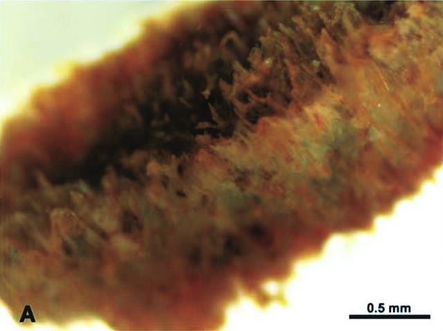
Wankel endosteal bone surface
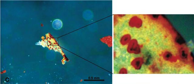
2) This is a photomicrograph of the Wankel rex again, showing tubular structures about 50 micrometers across, with the enclosed darker spheres. I'd love to have a medical student look at this without knowing what it was, just to hear what they said:

Wankel vessel
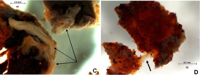
3) This is a shot of a piece from "Sue," showing the same type of micro-structures:
FMNH-PR-2081
Note that you can see darker centres to some of the red spheres in both of the above photomicrographs.
4) Here is a shot of a microstructure from the 2003 juvenile T. rex, MOR-1125, which shows internal structure, as well as thin extensions protruding from the main body:
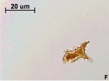
MOR-1125
This looks very much like an osteoblast or osteocyte, or a bone-producing cell, from any modern animal. The extensions are known as filipodia and they fit into thin channels called canaliculi that allow the cells to pass information and nutrients through the dense calcium phosphate (apatite) bone matrix (here is a slide showing osteocytes in place from a fossil theropod's toe!). While osteocytes have been seen before in fossils, we had never seen internal structure. The microstructures inside the 'osteocyte' in the picture above are interpreted as the remnants of cellular organelles. Schweitzer's team also found microstructures in the other two T. rexes and the hadrosaur Brachylophosaurus MOR-794 that looked very much like osteocytes. Immunoassay tests done against the materials from the first T. rex, MOR-1125, indicate that proteins from the original bone are highly likely to be present.
We do not know as much about what fossilization does and does not destroy as we thought we did. What Schweitzer's current work shows is that there are certain levels of physical microstructure that are preserved that we did not think were possible. Not much has yet been published about the possible preservation of the biochemical microstructure inside these things, which is where the holy grail of dino-DNA lies (but more on that later). It is slowly becoming clear that fossilization can preserve some very odd things - shapes of things certainly, like bones (even delicate ones in embryos), but also the overall shapes of soft tissues, like feathers, bananas, dinosaur heart, and even jellyfish. There are cases where fossil beetle casings and dinosaur feathers/hair (?) preserve their pigmentation, or colour.
But until now, no one had thought that cellular-level physical structures might be preserved. We knew that some of the original chemicals could be preserved - I worked with Heinz Lowenstam and Joe Kirschvink while I was at Caltech, and collected 63 million year-old Cretaceous ammonites (Baculites inornatus) from Baja California that still had mother of pearl lustre in them, in the unstable aragonite form (here are examples from Japan and from South Dakota). Based on ion exchange chromatography, Weiner, Lowenstam, Taborek and Hood found that the organic matrix in the shells of 80 million year old molluscs from Tennessee (Scabrotrigonia thoracica) probably had the primary, secondary and possibly even tertiary conformations of their proteins preserved. Weiner and Lowenstam also found that the isoleucine in fossil shells was sometimes not racemized - that is, it was still all of the same chirality (handedness), when most materials will naturally devolve into a 50-50 mix of epimers. The lack of epimerization for the shell in the intervening 63 million years, when the characteristic epimerization time is about 10^5 years, is thought to be due to the stabilization of the amino acids by the bioinorganic phase of the shell matrix. Whereas dinsoaur blood had never been found, traces of hemoglobin had been seen, by Schweitzer and others.
It exactly is this 'protection' afforded by the surrounding minerals in the dense bone that contributed to the preservation in the dinosaur soft tissues - as well as the burial under anoxic conditions in sediments that apparently did not allow bacterial action, or later flow of fluids that so often severely alter biological materials. The study of all this, how living materials become fossils, is called 'taphonomy,' and obviously these latest results will have a large impact on this field. One large point in the Schweitzer article is that these types of microstructures have never been found before not because they are rare, but because we had never thought them possible, and so had never even looked.
So the question really comes down to 'exactly what level of detail is preserved in these specimens, and can we expect even better examples?'
We can probably expect to learn a good deal about cellular structure, since the morphology seems to be well preserved, whether with the original biomolecules, or with substituted materials. If original biomolecules are present, or even their degradation products, then a lot can be learned - consider that the presence of certain proteins is in fact a flag for certain DNA sequences and metabolic pathways, so there can be a lot of work done on dino-DNA by inference. And this of course will be very closely watched by folks looking at the phylogeny of the dinosaurs - are modern birds really descendants of the theropods? Were the dinosaurs warm-blooded? Proteins will play a very large part in decyphering those stories.
It turns out I have an inside track on this one, since Mary Schweitzer, the scientist who found the soft tissue in the Tyrannosaurus rex bones, is working with funding from a paleontology program I co-direct. So I picked up the phone and called her - to congratulate her, and also to get an idea of what was ahead, now that the media was paying attention. She was also amazed at the media response, and had been on the telephone pretty much continuously for the whole week.
She is working on another publication, this time looking more closely at bone sub-types and physiological function as well as chemistry - 'nuff said. I had asked her for permission to use a particularly spectacular picture that was not in the Science article for this posting, but it is being used in the new article, so I will leave it for a later date. Although we did not speak about it, I can only assume that future work by Mary and others will explore whether dinosaur fossils have the types of cells associated with bone formation and maintenance (osteoblasts and osteocytes), as well as the cells that break down bone to supply calcium when needed (osteoclasts, for making egg shells, or bones in embryos).
In an interesting aside to all this, I saw that the discovery of well-preserved materials like this is always picked up by short-time creationists (1, 2, 3), as evidence that the Earth cannot be millions of years old. It's an interesting exercise to try and refute this argument, because both sides of Occam's razor are being used: it's a simple explanation (dino bones are only thousands of years old, not millions) and yet you would have to throw away a lot of other work to accept it (all the stratigraphy and geochronology). Here's one refutation. Then again, there are also long-time creationists in the fray...
Of course when I told my nine year old son about the dinosaur marrow, he came right back with: "Did you see the special the other night on Discovery Channel where they had found a real dragon?" After probing a bit, I think he knew the dragon was not real, but the fact that dragons had been mixed with dinosaurs on the TV show made him less apt to believe in the dinosaurs themselves - an interesting effect.
No comments:
Post a Comment